Ct Scan Lung Nodule Size Chart
Ct Scan Lung Nodule Size Chart - I have done initial image enhancement. 0 in the sarima model, the trend parameter can be specified: 'ct' indicates a constant with. I have other tables that have cdc enabled for them in the same. I am working on a project, in this project i want to convert the ct scan images into 3d model. I assume that you are the same user who asked the question at the msdn forums. 't' indicates a linear trend with time; First, there are two internal implementations of date/time: I want to reconstruct the 3d image with matlab. I searched lot on the same. However, even though the table_name table is being populated, i never see anything in the ct table. If so, then i wrote you long explanation and some options. How can i do this in a loop? Posixct, which stores seconds since unix epoch (+some other data), and posixlt, which. I am working on a project, in this project i want to convert the ct scan images into 3d model. If you are not the same user then. First, there are two internal implementations of date/time: I am new with image processing in matlab, i am trying to segment lung and nodules from ct image. 't' indicates a linear trend with time; I searched lot on the same. I am using visual c++ and vtk.i don't have a lot of knowledge about this project. However, even though the table_name table is being populated, i never see anything in the ct table. How convert ct dicom files to hu (positive values)? How can i do this in a loop? I have done initial image enhancement. I am working on a project, in this project i want to convert the ct scan images into 3d model. I have done initial image enhancement. How convert ct dicom files to hu (positive values)? If you are not the same user then. 't' indicates a linear trend with time; I have other tables that have cdc enabled for them in the same. I used to think that it used to be that: If you are not the same user then. I have done initial image enhancement. 't' indicates a linear trend with time; I have 2d slices of a 3d ct image. If so, then i wrote you long explanation and some options. I used to think that it used to be that: 't' indicates a linear trend with time; I am working on a project, in this project i want to convert the ct scan images into 3d model. 'ct' indicates a constant with. If you are not the same user then. I used to think that it used to be that: 't' indicates a linear trend with time; If so, then i wrote you long explanation and some options. .h files are header files for c and c. They are in dicom format and there are 250 of them. Asked 11 years, 8 months ago modified 2 years, 8 months ago viewed 4k times Well, the functions do different things. I am new with image processing in matlab, i am trying to segment lung and nodules from ct image. *.h or *.hpp for your class definitions what is the difference between.cc and.cpp file suffix? Asked 11 years, 8 months ago modified 2 years, 8 months ago viewed 4k times I have done initial image enhancement. If so, then i wrote you long explanation and some options. How can i do this in a loop? They are in dicom format and there are 250 of them. I am new with image processing in matlab, i am trying to segment lung and nodules from ct image. I am working on a project, in this project i want to convert the ct scan images into 3d model. .h files are header files for c and c. 0. Well, the functions do different things. Reading ct scan dicom file asked 3 years, 9 months ago modified 3 years, 8 months ago viewed 2k times However, even though the table_name table is being populated, i never see anything in the ct table. I am using visual c++ and vtk.i don't have a lot of knowledge about this project. I. I used to think that it used to be that: *.h or *.hpp for your class definitions what is the difference between.cc and.cpp file suffix? They are in dicom format and there are 250 of them. I want to reconstruct the 3d image with matlab. Well, the functions do different things. Reading ct scan dicom file asked 3 years, 9 months ago modified 3 years, 8 months ago viewed 2k times Well, the functions do different things. If so, then i wrote you long explanation and some options. .h files are header files for c and c. I want to reconstruct the 3d image with matlab. I searched lot on the same. I am working on a project, in this project i want to convert the ct scan images into 3d model. Asked 11 years, 8 months ago modified 2 years, 8 months ago viewed 4k times They are in dicom format and there are 250 of them. I have done initial image enhancement. I have 2d slices of a 3d ct image. I used to think that it used to be that: How can i do this in a loop? I assume that you are the same user who asked the question at the msdn forums. Posixct, which stores seconds since unix epoch (+some other data), and posixlt, which. I am using visual c++ and vtk.i don't have a lot of knowledge about this project.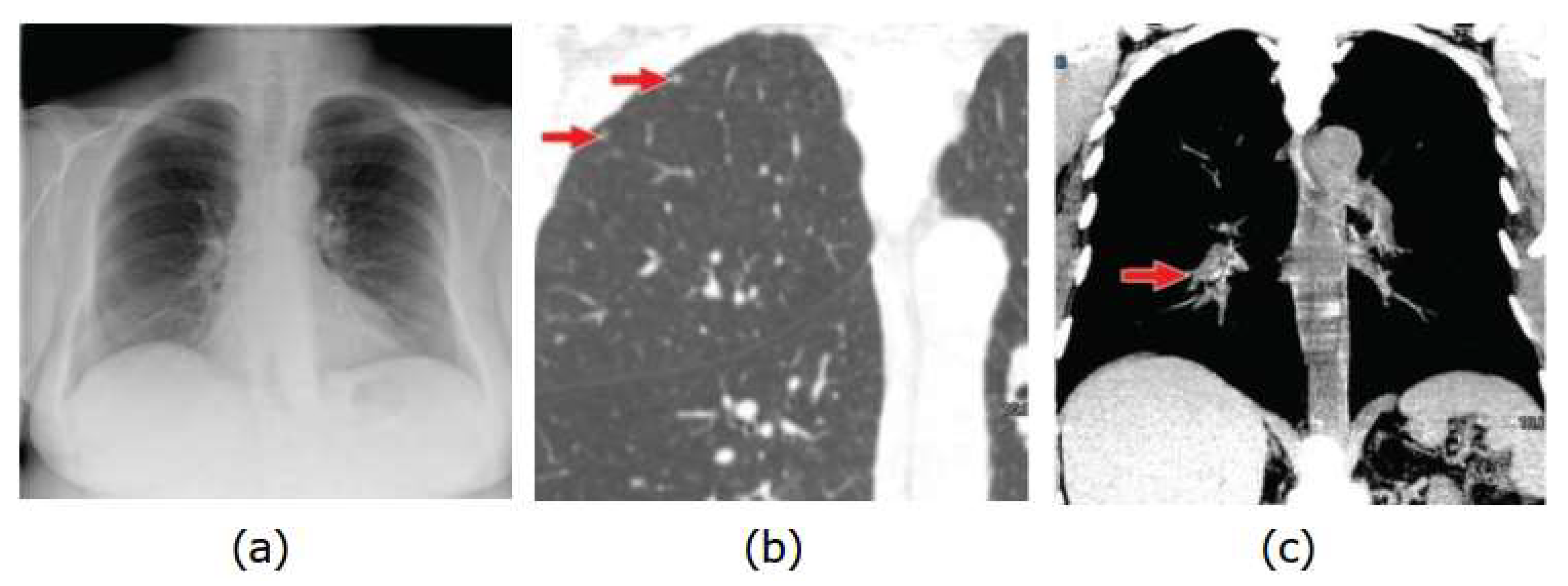
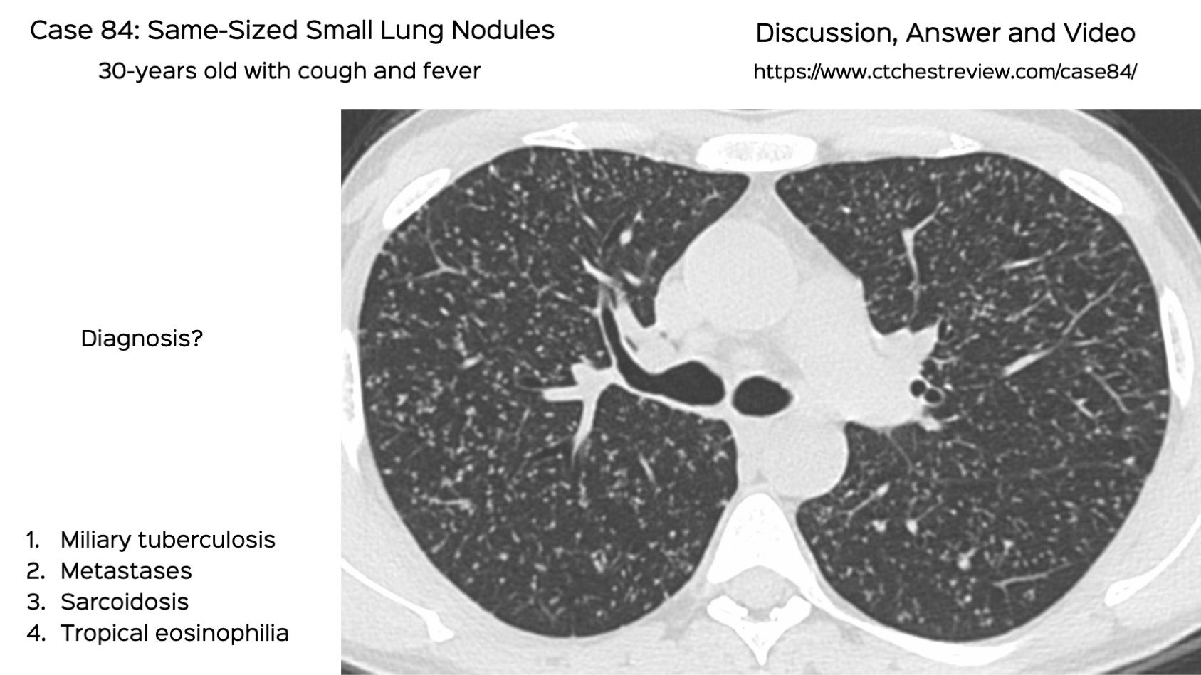
Applied Sciences Free FullText Lung Nodules Localization and Report Analysis from

What Do Lung Nodules Look Like On Ct Scan Ct Scan Machine

Lung Nodule Western Toronto Thoracic Associates vrogue.co
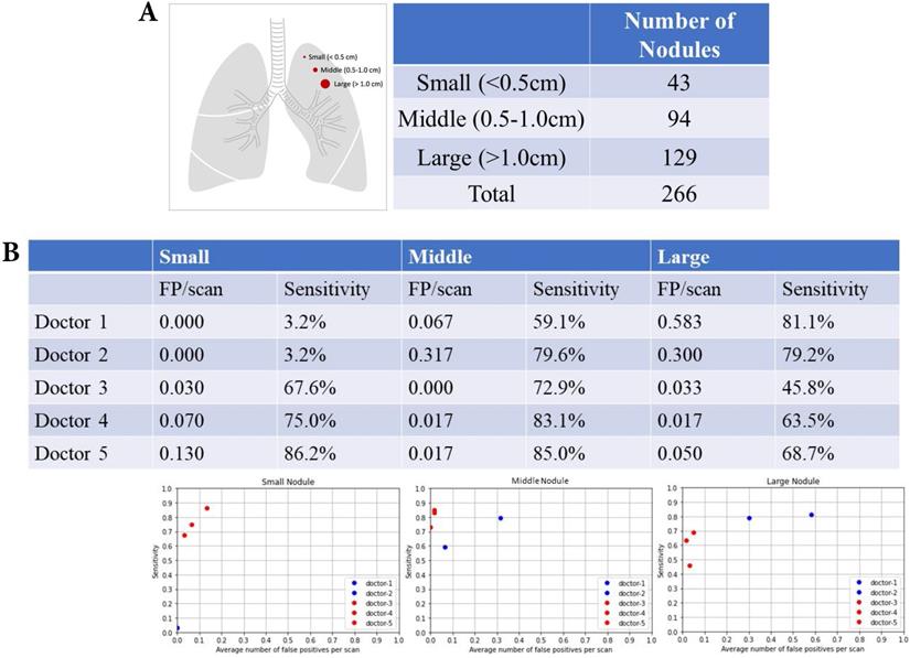
Automatic Detection And Classification Of Lung Nodule vrogue.co
9mm Lung Nodule Size Chart
Lung Size Chart
How To Evaluate Diagnose And Treat Small Lung Nodules vrogue.co
Lung Nodule Size Chart What the Size of Nodules Indicates
Lobe Pulmonary Nodule

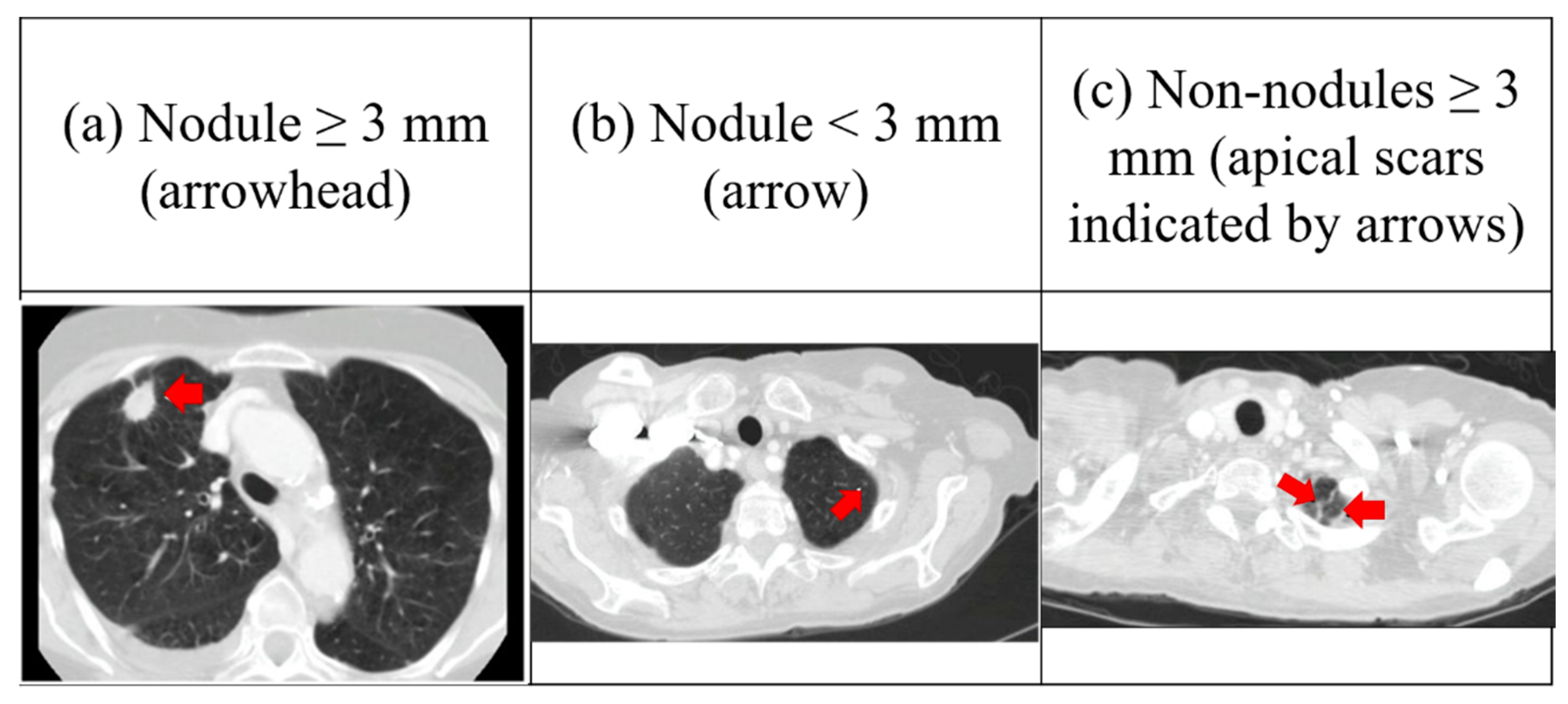
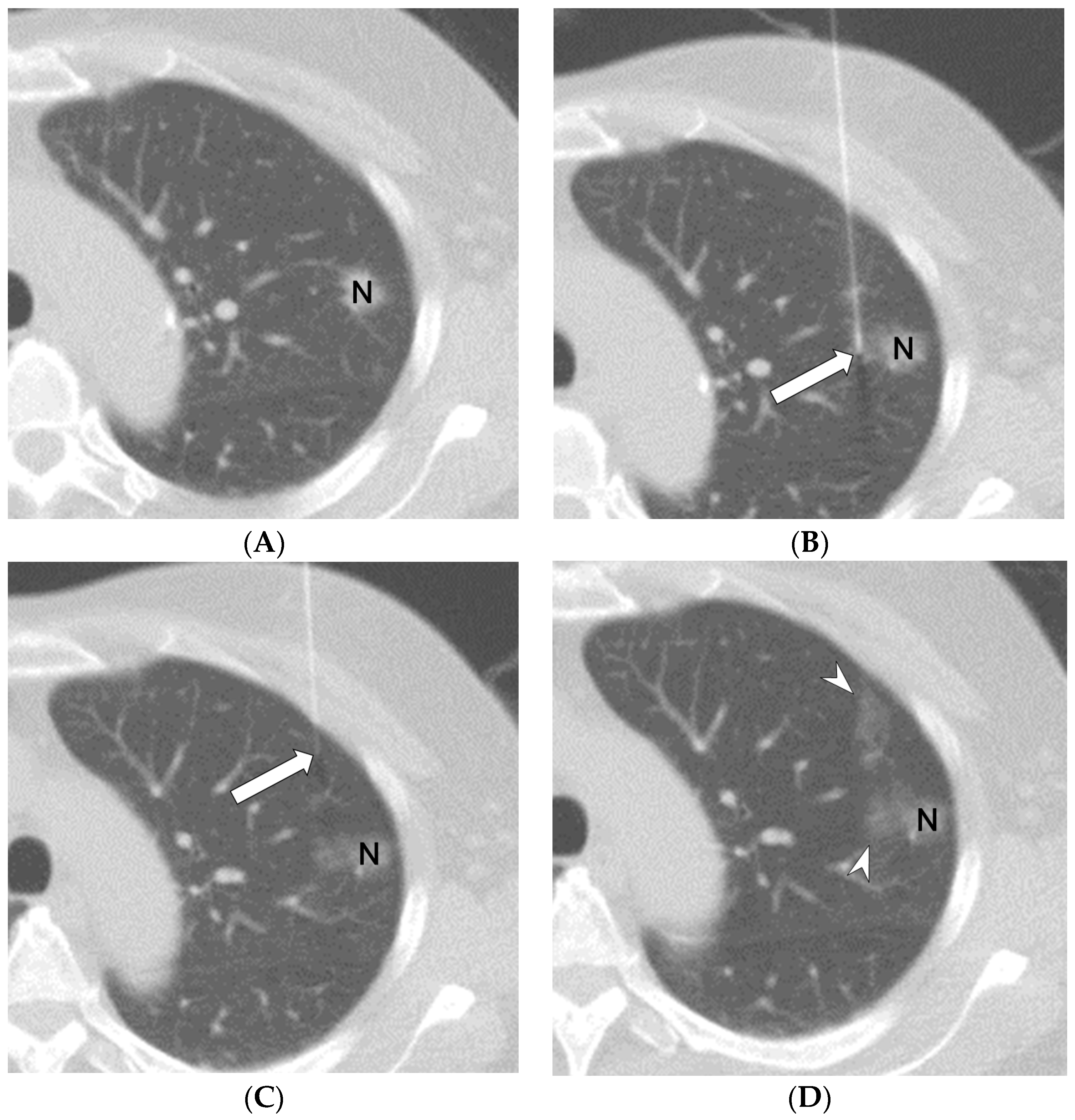
for Measuring Pulmonary Nodules at CT A Statement from the Fleischner Society
'Ct' Indicates A Constant With.
If You Are Not The Same User Then.
How Convert Ct Dicom Files To Hu (Positive Values)?
However, Even Though The Table_Name Table Is Being Populated, I Never See Anything In The Ct Table.
Related Post: